
Our models:
advantages
and properties
Silk4B’s patented in vitro silk-based 3D bone marrow models reproduce faithfully the properties of the in vivo bone marrow niche. The models enable in vitro studies with high translational relevance to human biology, while offering ease-of-use, scale, and cost-effectiveness.
The bone marrow is one of the most dynamic and complex body organs, highly dependent on several factors to ensure proper function. Biochemical signalling and cellular interactions ensure correct cell proliferation, survival, and differentiation of cells, as in many other tissues. However, in contrast to other tissues, the matrix architecture and its biomechanical properties are not just a physical frame, but are essential for proper bone marrow cell survival and function. In many diseases, changes in these biomechanical properties are a hallmark.
Silk4B’s patented in vitro 3D bone marrow models are based on silk fibroin, a silk protein, and for the first time recreate the physical properties and matrix architecture of the bone marrow in vitro. Silk fibroin was chosen as the core material because, unlike many other biomaterials, it does not activate platelets, a critical property for building a bone marrow model capable of reliably studying hematopoiesis without introducing artificial bias. Our models allow proper biochemical signalling and cellular interactions. Altogether this ensures that survival, proliferation and differentiation of bone marrow and blood cells of interest is mimicking closely the in vivo bone marrow structure, and the experimental results using our models have high translational efficiency.
Section 1Biological relevance with high translational value
Due to faithful reproduction of the bone marrow structure, our 3D bone marrow models allow for:
increased cell survival and functional maintenance
successful differentiation into functional mature cell types
(e.g., platelets, red blood cells).
This ensures translationally relevant studies of hematopoiesis and bone marrow and blood cell function.
Tunable structure complexity
Our 3D bone marrow models work successfully when using a single cell type or a co-culture of multiple cell types. This allows to dissect complex processes in normal biology and disease.
Home› Our models › Advantages & Silk Properties
Section 2
Disease relevance and targeted therapy development
Our 3D bone marrow models allow to dissect disease biology and identify relevant therapy targets for drug development:
Disease contribution of physical environment vs cell intrinsic properties: Our models come with tunable physical properties allowing to model the biomechanics of normal and diseased environment, and combined with seeding of normal or diseased cells, this allows to dissect the contribution of physical environment and cell intrinsic properties on disease phenotype and progression
Disease contribution of specific cell types: By allowing multiple cell type co-culture our models allow to combine normal and diseased cells to identify which cell types and cell-type specific molecular pathways play a role in disease.
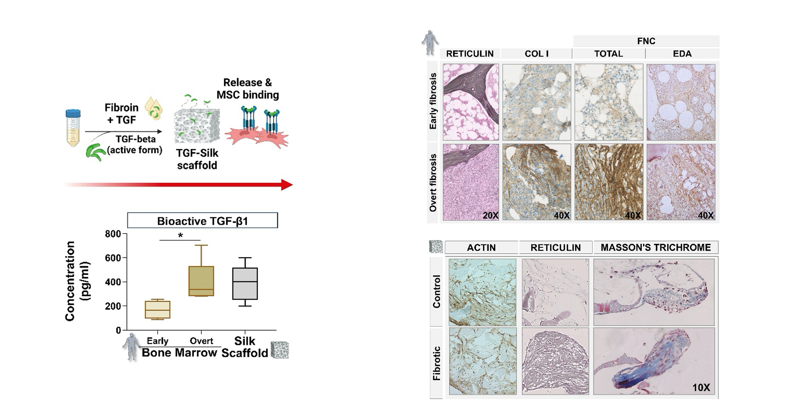
TGF-beta1-functionalized silk scaffolds reproduce a fibrotic bone marrow microenvironment. Schematic of the TGF-beta1 functionalization process: recombinant TGF-beta1 is loaded into silk fibroin scaffolds, where it is stabilized and subsequently released at concentrations similar to those measured in patients with overt fibrosis. Representative histological images of bone marrow biopsies from patients with early and overt fibrosis showing progressive extracellular matrix (ECM) accumulation. Histological analysis of scaffolds and Masson’s trichrome reveals increased fibrosis-like ECM deposition in TGF-beta1-loaded constructs (magnification: 10×). Small 2026
Section 3
Fits existing workflows of low to high throughput capacity and automation
Our 3D models come in two forms (hydrogels and scaffolds) to meet the needs of varied experimental designs allowing for a range of experimental complexity and throughput. Furthermore, our SilkInk™ hydrogel was specifically designed to work with 3D bioprinters and easily fit into existing workflows that involve automation.
High mature cell yield per experiment
By faithfully recapitulating the bone marrow structure and biologically relevant cell behaviour, cell differentiation in our 3D models is not only biologically relevant leading to terminally differentiated cells but also high yield. When comparing to standard in vitro 2D liquid culture, the yield of differentiated cells from the same amount of progenitor cells is several fold higher in our model (e.g., 5-6 fold higher yield in differentiated platelets.)
Time and Reagents Savings
While delivering superior biologically translatable experiments and higher yield of terminally differentiated cells compared to 2D liquid culture, our models also require fewer reagents for the same amount of progenitor cells and less hands-on personnel time for handling compared to standard 2D liquid culture. For example, platelet differentiation from amount of progenitors in our models results consumes 70-80% fewer reagents and takes 60-70% less hans-on personnel time than 2D liquid culture.
Works with multiple cell sources, including sensitive and valuable patient-derived cells
Our 3D models work with blood and bone marrow cells from multiple sources, including patient HSPCs, peripheral blood cells, bone marrow biopsy cells, umbilical cord blood cells, cell lines, and iPSC derived cells
Works with multiple species
Our 3D models work with cells from humans and mice, the two most relevant species for research and drug development.
Easy to setup and use
Our 3D models are as easy to set up as standard in vitro 2D liquid culture. The process is technically simple for seeding and extracting cells from our 3D matrix. Standardized protocols are available for culturing, proliferation and differentiation of various cell types in single or multiple cell type culture.
Easy microscopy
Our 3D models have high transparency, making microscopy easy and enabling live imaging and staining.
Validated in peer-reviewed published research and trusted by academic and pharma customers
Our 3D models have been validated in multiple [>30] peer-reviewed studies and are being used by academics and pharma companies for research and drug discovery.
Silk Properties
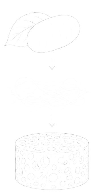
All of this is possible due to our patented approach of using Silk Fibroin as a fundamental material in our 3D bone marrow models. Silk Fibroin has unique properties that make it suitable for fundamental and disease relevant research as well as medicine.
Tunable stiffness: Silk Fibroin has the unique property that it can be processed to create different physical stiffness levels for our 3D bone marrow models. The very soft versions (SilkInk, SilkThemo, SilkFold) are the only in vitro models in research that are matching the healthy bone marrow structure. Stiffer models (SilkTune with its tunable stiffness) allow to match the stiffness of diseased bone marrow structures. This makes our models unique in their ability to allow dissecting the physical effects of the bone marrow environment and disease progression.
Biocompatible: Silk Fibroin is a highly biocompatible material known for its low immunogenicity, non-thrombogenic properties, acellular and no xenogenic risk. This makes it suitable for work with delicate cells and even patients and it has been approved by FDA and EMA for medical use.
3D architecture: Silk Fibroin helps maintain the 3D architecture in our models allowing for proper cell-matrix and cell-cell interactions
Molecular biocompatibility: Silk Fibroin allows for delivery of molecules to the cells (cytokines, growth factors, other). This is an essential property that allows the necessary molecules to reach the cells for their survival, proliferation and differentiation in our 3D models.
Optical transparency: Silk Fibroin is highly transparent optically, which makes it suitable for microscopy, including live imaging.