
Erythropoiesis in silk-based 3D bone marrow models
Applications
Silk4B’s 3D bone marrow models allow for enucleated erythrocyte differentiation and collection.
Home › Applications › Erythropoiesis
In vivo mimicking erythropoiesis using in vitro 3D bone marrow

In vivo-like differentiation into erythroblastic islands (EBIs) and enucleated erythrocytes (red blood cells, RBCs)
Easy erythrocyte collection
Silk4B’s silk-based 3D bone marrow models allow differentiation into enucleated erythrocytes, or red blood cells (RBCs), that resemble peripheral RBCs found in the human body.
The differentiation process mimics in vivo steps with sequential differentiation into erythroblastic islands (EBIs). The morphology of erythroblastic islands in our models resembles that of in vivo EBIs. The EBIs in our models contain a gradient of less mature CD71+ CD235- cells to more mature CD71low CD235+ cells. Transmission electron microscopy clearly identifies cells from progenitor ProE cells to Rtics. Eventually, enucleated biconcave RBCs form at the periphery of EBIs. Collected RBCs are primarily enucleated and are similar to peripheral RBCs, they show very few residual mitochondria and glycogen granules.
RBCs can easily be isolated and used in downstream assays. RBCs collection is easy from both silk hydrogel and silk scaffold models. Scaffold models allow for perfusion of the model with platelets released into the perfuate mimicking the RBC release into the blood. The release of RBCs into the perfuate allows their easy collection.
When compared to 2D liquid culture models, RBC differentiation in our model results in differentiation of significantly more mature cells that are enucleated. This is supported by comparative transcriptomic analyses which show that unlike 2D liquid culture, our 3D bone marrow models support autophagy processes which promote organelle clearance and membrane remodelling. Importantly, in 2D culture cells experience significant stress that causes increased gamma-globin production; while in our 3D models cells show less gamma-globin production due to lower stress.
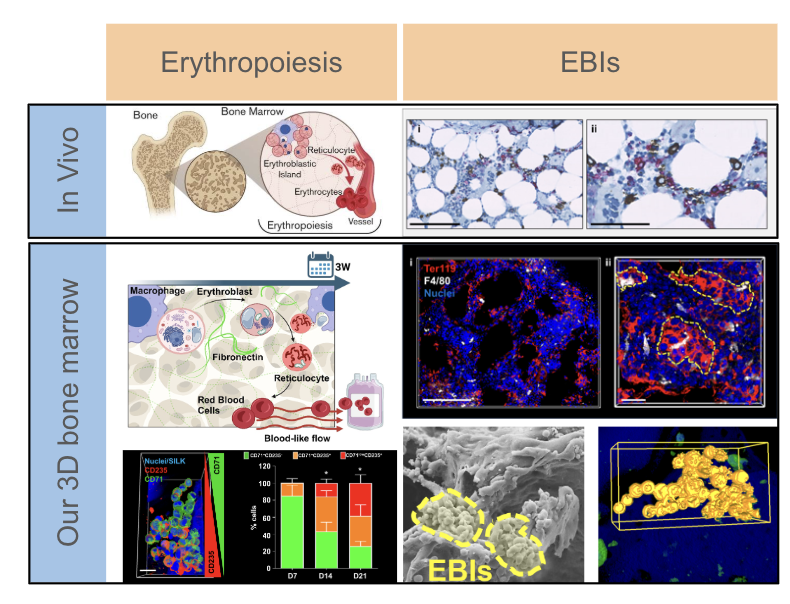
Schematic outlines of erythropoiesis in vivo and in vitro in Silk4B’s 3D bone marrow scaffold model.
Bottom left: In our scaffold model, cells form EBIs that have proportionally more mature cells over the course of differentiation (day 7,14,21) and show a gradient of less to more mature cells within the islands.
Right: EBIs in vivo and in vitro in our model
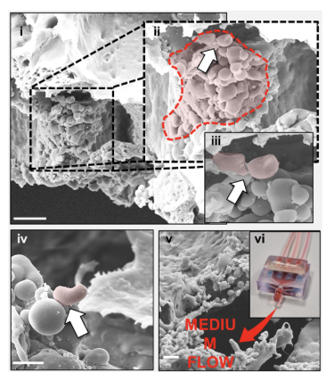
SEM of scaffold model showing the presence of biconcave enucleated RBCs (i-iv), at the periphery of EBIs (white arrows indicate biconcave RBCs)
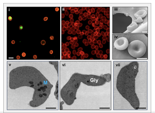
RBCs from our scaffold model (i) have been imaged by epifluorescence microscopy (red, CD235; green, nuclei; scale bar, 5 μm); peripheral blood RBCs have been used as reference control (ii); SEM analysis (iii-iv) of collected RBCs; cells show morphological features of native RBCs, including the biconcave discoidal shape; transmission electron microscopy (v-vii) of collected RBCs. Enucleated cells show few small mitochondria and some residual glycogen deposits. Gly, glycogen; M, mitochondria.
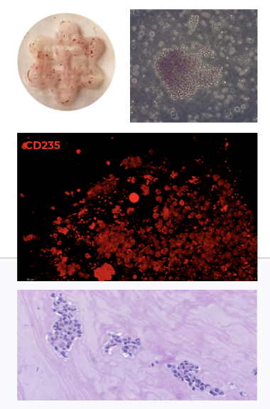
Silk Hydrogel with differentiated CD235 positive cells inside. Bottom: H/E staining of hydrogels and cells inside