
Applications
Disease modelling
Silk4B’s 3D bone marrow models allow in vitro culturing of malignant cells, mimicking of the physical properties of a malignant environment, and dissecting the contribution to disease of intracellular, cell-cell and cell-matrix interactions.
Home› Applications› Disease modelling
Leukemia
Our 3D bone marrow models are suitable for growing and studying leukemic cells. When our 3D bone marrow scaffolds, pre-seeded with MSCs, are perfused with leukemic cells (PLB-985), the leukemic cells attach to the bone marrow model (video left).
This attachment is not purely physical, as cells can stay attached for hours while they are continuously perfused with media. As cells proliferate, some of the cells will detach from their cluster and enter again the flow (video right), mimicking malignant cell detachment and entering blood flow in vivo. This allows investigations into why cells lose their attachment; helping to understand disease spread mechanisms and identifying potential drug targets for therapy.
Myeloproliferative Neoplasms
Our 3D bone marrow models are suitable for studying myeloproliferative neoplasms (MPNs) and dissecting how intracellular properties, cell-cell interactions and matrix properties affect disease phenotype.
Intracellular properties in MPNs. Megakaryocytes (MKs) from JAK2V617F MPN patients show progressive cell surface area reduction as bone marrow fibrosis progresses in these patients. The specific impact of the niche on malignant megakaryocyte (MK) behavior, including proliferation and maturation, remains poorly understood. Using our models, it was possible to investigate whether MK cell surface reduction is impacted by the physical changes in the bone marrow. MKs isolated from JAK2V617F patients and cultured in our 3D bone marrow scaffold with stiffness properties mimicking healthy bone marrow had a smaller surface area compared to healthy cells in a healthy niche, indicating that cell-intrinsic properties affect their phenotype.
Matrix properties in MPNs. MKs from JAK2V617F patients in the normal in vitro 3D bone marrow silk scaffold had a higher cell surface area compared to those in the fibrotic-mimicking in vitro 3D bone marrow niche that has stiffer properties. This showed the additional effect that cell-matrix interactions have on the disease phenotype. These stiffness effects translate to platelet production, as even healthy MKs derived from HSPCs show a lower percentage of proplatelet formation, and overall fewer platelets are made when they are placed in the fibrotic-mimicking 3D bone marrow silk scaffold with high stiffness compared to the normal 3D bone marrow. Using our products with diverse stiffness levels researchers can model malignancies where the physical properties of the bone marrow niche are known to be changing in vivo during disease progression.
Cell-cell interactions in MPNs. Our 3D bone marrow models are also suitable for co-culture of multiple cell types and allow dissecting the contribution of specific cell types and cell-cell interactions to disease pathology. Studies in our 3D bone marrow models using healthy and diseased megakaryocytes and neutrophils from patients with JAK2V617F myeloproliferative neoplasms confirmed defective neutrophil clearance via the immune checkpoint CD24 as a cause of the disease.
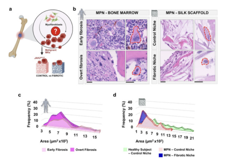
(a) Schematic illustration of MPN‐associated bone marrow fibrosis: JAK2V617F malignant MKs have been cultured within the two distinct 3D bone marrow silk scaffolds: control niche (non‐fibrotic) and fibrotic niche.
(b) Bone Marrow: Histological analysis of bone marrow sections from patients with MPN, showing MK morphology in early versus overt fibrosis stages. Silk scaffold: Histological analysis of control versus fibrotic silk scaffolds cultured with MPN‐derived MKs (hematoxylin and eosin staining).
(c) Frequency distribution of megakaryocyte surface area in early (light pink) and overt (dark pink) fibrotic stages, showing a shift toward smaller megakaryocytes in overt fibrosis.
(d) Frequency distribution of megakaryocyte area from healthy subject samples (green) cultured in the control niche, MPN samples cultured in control (red) versus fibrotic (blue) niches, confirming a fibrotic niche‐induced shift toward smaller megakaryocytes.