Applications
Cell maintenance
and function studies
Silk4B's 3D bone marrow models maintain and proliferate multiple bone marrow cell types, alone or in co-culture, and support multi-lineage differentiation into mature, functional blood cells
Home › Applications › Cell maintenance and function studies
Cell maintenance and proliferation
The in vivo bone marrow niche involves close contact and communication between the matrix and multiple cell types to regulate blood cell production. Silk4B's silk-based 3D bone marrow models are built to support this cellular diversity, allowing to grow individual cell types or co-cultures and working with a variety of cells, from megakaryocytes, to neutrophils, to stromal cells, to endothelial cells, and hematopoietic stem cell progenitors (HSPCs). Our models work with primary cells from mobilized peripheral blood, cord blood, or bone marrow biopsy, as well as differentiated cells from iPSCs and also cell lines.
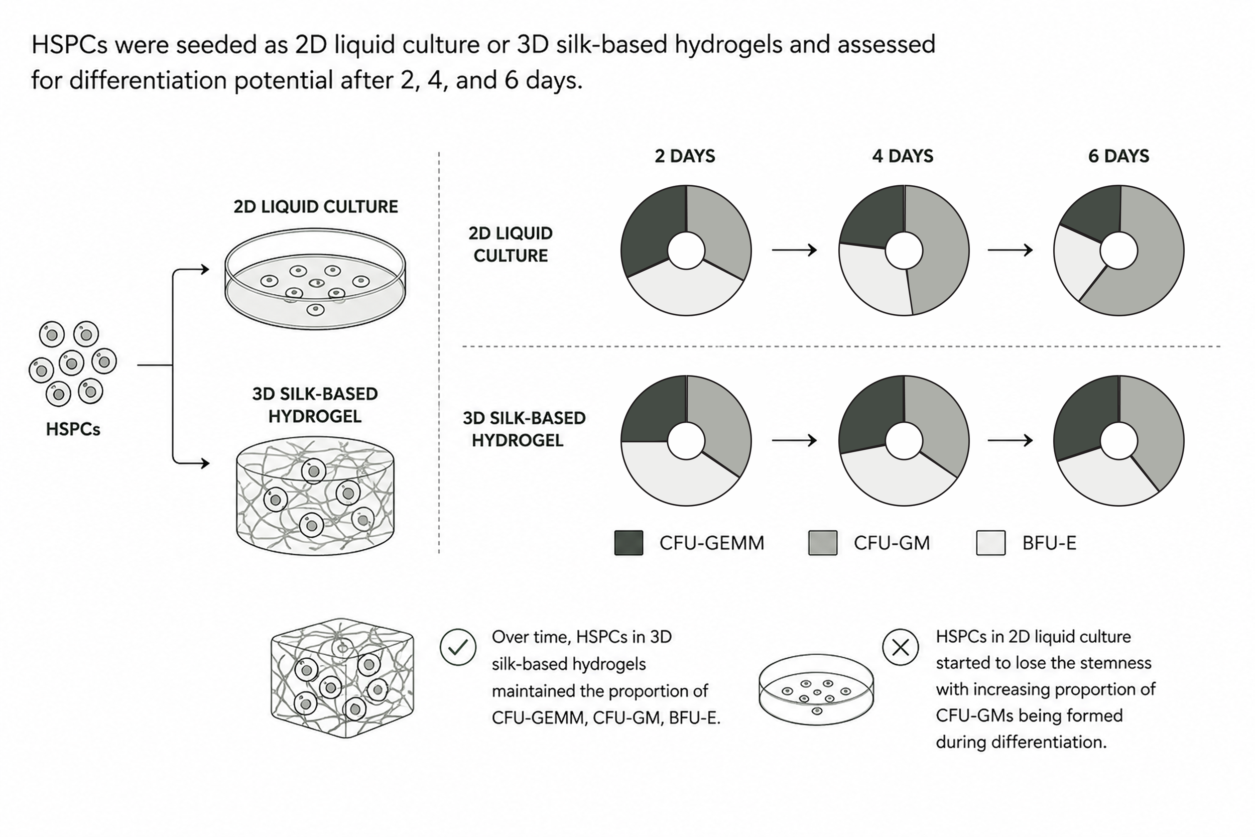
Maintenance of cells in our models is superior to 2D culture and other standard preservation methods. Peripheral blood cells, for example, neutrophils show better survival (fewer apoptotic cells over time) compared to standard pellet preservation, and remain functional (participate in megakaryocyte emperipolesis when co-cultured in our 3D bone marrow models). HSPCs also show high survival lower chronic cellular stress than in 2D liquid culture (where they upregulate pathways linked to replication stress, DNA damage response, and redox stress) that distort cell identity over time.
The more natural matrix environment and lower stress allow the HSPCs proliferating in our in vitro 3D bone marrow models to retain their stemness and multi-lineage differentiation potential over time, unlike in 2D liquid culture where over time cells drift towards preferential CFU-GM lineage during differentiation. These healthy cells with preserved, multipotent progenitor potential lead to more in vivo mimicking differentiation processes and mature cell outcomes.
Cell differentiation
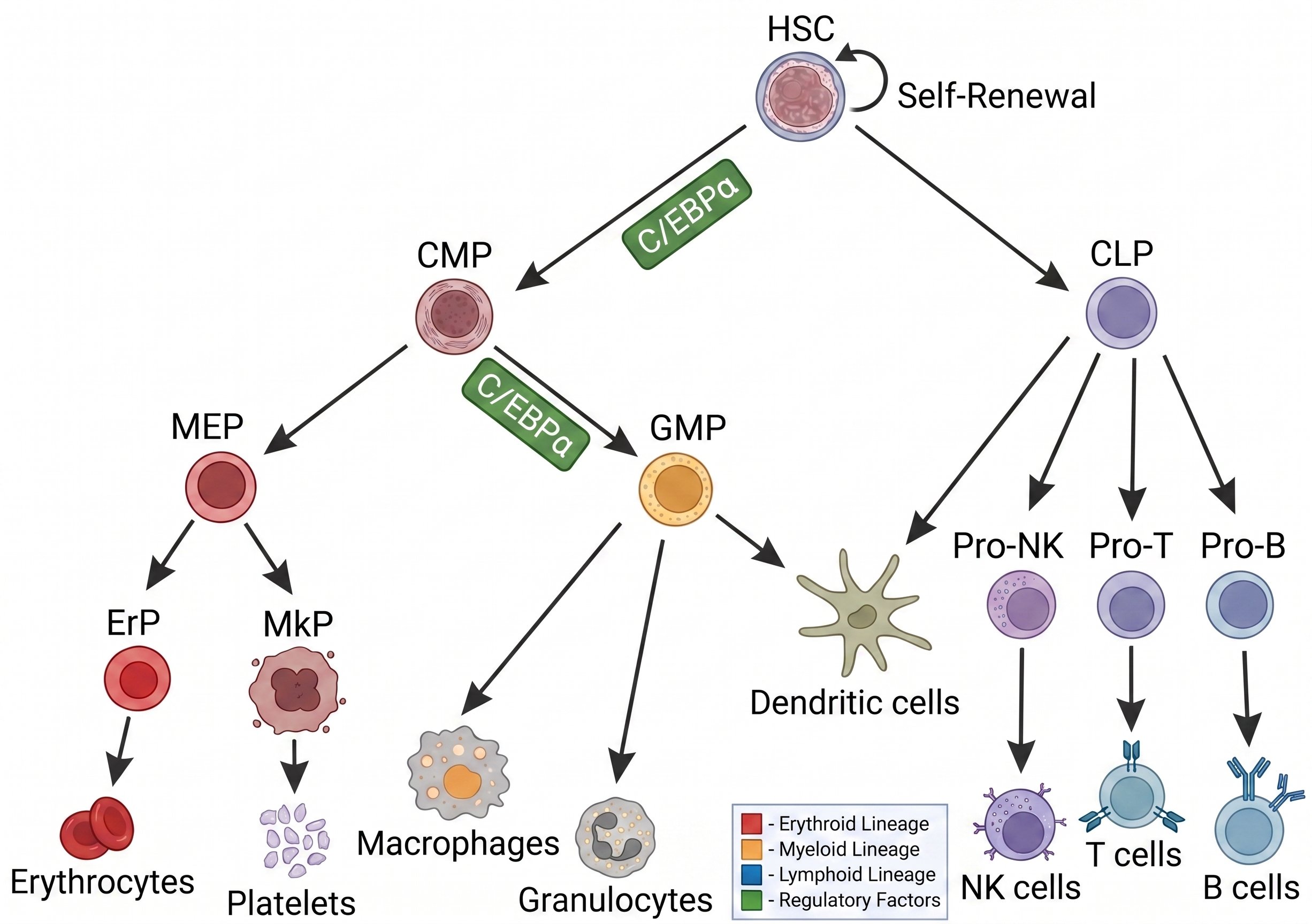
As our in vitro 3D models provide an in vivo mimicking environment that maintains HSPCs to be healthy and retain their full differentiation potential they can be directed to different lineages and the cells along that lineage are maintained in the right bone marrow like matrix allowing for creation of mature, functional blood cell types that resemble the in vivo peripheral cells. This has been established extensively with peer reviewed and highly reproducible protocols for two lineages: thrombopoiesis (platelets) and erythropoiesis (red blood cells).
Thrombopoiesis (platelets)
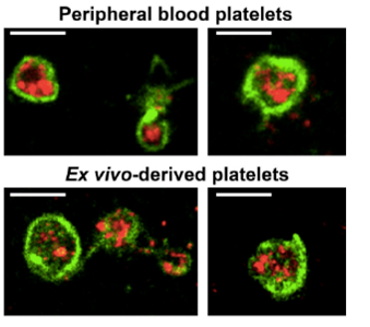
Our models support differentiation into mature, functional platelets from primary HSPCs, iPSC-derived progenitors, or primary megakaryocytes and work with human or mouse cells. The process follows the in vivo progression from early progenitors through late progenitors to megakaryocytes. The models are non-thrombogenic, so platelets do not aggregate and can be isolated for downstream assays. In the 3D scaffold bone marrow format, when perfusion is used, platelets are released into the perfusate allowing easy collection and studying platelet release.
Differentiation is considerably more effective than in 2D: from the same number of starting progenitor cells, our models yield roughly 6x more platelets than 2D culture or other conventional bioinks. Differentiation outcomes can be further modulated through co-culture. HSPC with stromal (MSC) support cells, or megakaryocytes with endothelial cells show further increased mature platelet production. Neutrophil and megakaryocyte co-culture, also increases platelet production via emperipolesis. These co-culture interactions can be dissected at the molecular level.
The 3D architecture and its in vivo mimicking effect on cells also reduces reagent use (~80% lower reagent cost for the same input progenitor cell number) and cuts personnel hands-on time for a full differentiation protocol from about 3 hours to roughly 0.9 hours (~70% savings) compared to 2D culture due simplifies media changes, since cells remain within the matrix rather than requiring collection and centrifugation.
Erythropoiesis (red blood cells, RBCs)
Differentiation involves co-culture with macrophages and mimics the in vivo sequence of erythropoiesis, forming erythroblastic islands (EBIs) evidenced by electron microcopy as well as immunohistochemistry showing a maturation gradient from less mature CD71+CD235- cells to more mature CD71low CD235+ cells, consistent with EBI morphology in vivo. Collected cells are primarily enucleated, biconcave RBCs with very few residual mitochondria or glycogen granules, similar to peripheral RBCs.
As with platelets, RBCs can be collected from either the hydrogel or scaffold format, with scaffold perfusion mimicking RBC release into the bloodstream.